These are generally regarded as deformities that do not threaten life. Because scoliosis (curvature of the spine) accompanies them in about one in four patients, this should be investigated. Patients should also be evaluated for various congenital heart diseases and other conditions.
Pectus Excavatum
Also known as funnel chest or sunken chest (cobbler's chest). It is an inward depression of the rib cage.
It is the most common of the chest wall deformities, occurring in about 1 in 300-400 live births. It is not as rare as is commonly thought. These children have a very typical appearance: a narrow and thin chest, rounded shoulders, flared ribs and poor posture. Pectus excavatum usually appears at birth or in the first years of life and is noticed by the mother and relatives. Although it very rarely regresses, the depression will most likely worsen during puberty. It is seen 4 times more often in males than in females. Although the cause of the depression is overgrowth of the costal cartilages, the exact etiology is not known. The irregularly growing costal cartilage pushes the breastbone backward. There is an increased familial predisposition: 37% of cases have a family history of chest depression (pectus excavatum).
In childhood this deformity is tolerated. As the patient grows older, pain in the deformed costal cartilage and pain in the left breast area may occur after exertion. Palpitations and heart rhythm disturbances may be observed after exertion. A heart murmur may be heard after short exercise. Electrocardiographic changes may occur because of displacement of the heart. When the chest wall deformity is corrected, an improvement in lung function is observed. With delayed corrections (after the age of 40), this improvement is somewhat harder to achieve.
The backward displacement of the chest wall causes a dent, particularly in the anterior wall of the right ventricle. When the depression is corrected, the dent in the ventricle also resolves. Put simply, the volume of the depression is space stolen from the heart and lungs. In my own words, I always warn my patients: "This deformity will cause you problems not in youth, but in old age." In old age, the heart and lung capacity stolen by the deformity will be needed more. In addition, deformity-related postural problems will lead to pain and limited mobility in old age.
For respiratory and cardiac problems, the greater the depression before surgery, the greater the improvement that should be expected. Many researchers have attributed the symptomatic impairment in pectus excavatum to a reduction in volume within the chest cavity. Because there is a wide range of cardiopulmonary function limits even in normal individuals, this relationship is difficult to prove. It depends entirely on physical activity and body habits.
Moreover, even if it causes no functional impairment, the patient's unhappiness with the deformity is in itself a reason for correction. The literature accepts this as well. Indeed, there are quite a few patients who say they never go into the sea without keeping their T-shirt on, expressing their unhappiness this way. Since 1960, psychiatrists have carried out many studies on this subject and have used the term "psychological deformity" for this condition. In one study, problems such as anxiety, loss of motivation and fear of being labeled (82%), lack of self-confidence and shyness (78%), indecisiveness (72%) and aggression (66%) were identified.
Treatment
There are both surgical and non-surgical treatment options for chest depression.
Two surgical methods are most commonly used. We will try to describe the Nuss (MIRPE - Minimally Invasive Repair of Pectus Excavatum) and Ravitch procedures.
In the Ravitch procedure, the chest is opened along its entire length and the deformed costal cartilages, except for the 2nd rib, are removed. A wedge-shaped piece is then removed from behind the most depressed part of the breastbone and closed with wire sutures. The procedure is completed by placing a bar behind the breastbone. Unfortunately, this operation is not as short as it sounds: it takes about 3 hours, involves bleeding, and 2 drains are placed to evacuate the blood. The bar is removed after about 10 days. The hospital stay and the time to return to normal life are long. The patient cannot play sports for at least 6 months. For these reasons it has lost popularity today (especially over the last 15 years). However, it can still be used as an option for deformities that cannot be corrected with the minimally invasive method.
As another method, raising the breastbone with a bar placed behind it has been reported. The minimally invasive procedure now known as the Nuss procedure has been developed and perfected over the years. Complications of this minimally invasive procedure are quite rare. The recovery period averages 4-5 days. Attention is paid to postoperative pain control, respiratory physiotherapy, and patient/parent education. The pectus bar is removed two to four years after it is placed. The procedure is performed under general anesthesia. In long-term results, the recurrence rate averages below 5%. If the deformity can be corrected with a single bar, this method is performed through a total of 3 surgical incisions of 2 cm each, 1 on the left and 2 on the right. Besides all these advantages, although very rarely observed, cardiac injury has been reported during insertion and sometimes removal of the bar with this method.
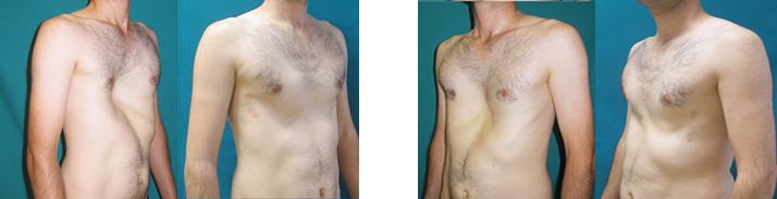
Image of one of our patients corrected with the Nuss method. The incision site is almost invisible.
There is also an alternative correction method free of these life-threatening risks: silicone implant placement. See here for details.
The non-surgical treatment is the application of a vacuum bell. In individuals up to 14-17 years of age, while bone development and hardening have not yet occurred, negative pressure is applied to the deformity daily with the help of a vacuum bell, and the deformity improves in about 1 year. It may not be suitable for wide oval-type defects or plateau-type defects. The right treatment is determined by the physician together with the patient. In addition to vacuum bell treatment, exercises performed with a physiotherapist and nutritional support provided with a dietitian are of great benefit in helping patients correct their deformities faster and more effectively.
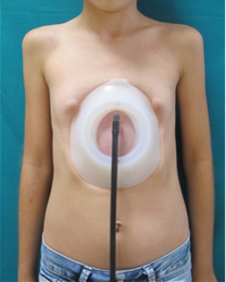
Image of a patient using a vacuum bell
Pectus Carinatum
Deformities in which the chest wall is not sunken inward but protrudes outward are called pectus carinatum. This deformity is much rarer than pectus excavatum. It does not usually cause a decline in heart or lung function. The main reason for treatment is that the patient is unhappy with this appearance.
Treatment
Its treatment is surgical, just as with pectus excavatum. Likewise, with the minimally invasive MIRPC (Minimally Invasive Repair of Pectus Carinatum) method (Abramson procedure), a steel bar placed under the skin can press the chest wall down to its normal position; alternatively, with the Ravitch procedure, a large incision is made, the sternum is detached from the cartilage and, if necessary, the cartilage is shortened. The difference between the Abramson procedure and the Nuss procedure applied to pectus excavatum patients is that the chest cavity is not entered; the bar is passed over the bone. For this reason, serious complications such as cardiac injury are not observed.
Below are the before and after images of a patient we operated on with the Abramson method.
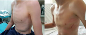
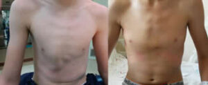
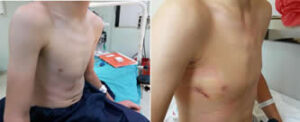
Below is the before and after appearance of a pectus carinatum case corrected with the Ravitch procedure.
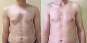
In non-surgical treatment, the use of a brace (orthosis) stands out. By applying continuous pressure to the protruding area with the brace, the deformity improves as the other parts develop. For brace treatment, there are approaches in which it is worn 23 hours, 18 hours, 12 hours or 6 hours a day. The longer the daily duration chosen, the sooner results can be obtained. However, this depends on the patient's social life. Selecting a suitable brace, making sure it is made of suitable material, and the need for close follow-up of the patient so that pectus excavatum does not develop while the brace is used make it appropriate to apply this treatment in experienced centers that perform it in large numbers. Patients should be followed with periodic examinations to determine how long treatment will continue and, if necessary, whether use can be paused from time to time and treatment repeated until the bones harden.
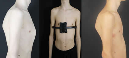
Appearance of a patient treated with a brace for pectus carinatum, before, during and after 10 months of treatment
For appropriate patient selection for brace treatment, the chest wall must not have hardened yet. For this reason it is applied to patients under 17 years of age. The pressure at which the deformity corrects should be measured, and patients whose pressure level is above a certain value should be offered surgery instead of a brace. In addition, at follow-up visits with pressure measurement, even when no result is yet visible in the patient's appearance, if the pressure required for correction has decreased compared with the value measured at the previous visit, it can be determined that treatment is on the right track and that a visual result can also be achieved if treatment continues. This prevents unnecessarily abandoning treatment.
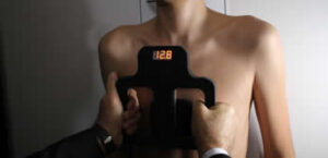
Measurement of the correction pressure in a patient with pectus carinatum
Poland Syndrome
It is quite rare compared with pectus excavatum and carinatum deformities. In people with this syndrome, one or several of the following signs and symptoms may be seen:
• Small or absent breast tissue or nipple
• Underdevelopment or absence of the chest wall muscles on the affected side
• Underdeveloped subcutaneous fat tissue
• Absence of underarm hair on the affected side
• Thoracic deformity due to absent or underdeveloped ribs
• Short arm or forearm on the affected side, shortness of the fingers (brachydactyly) and fusion of the fingers (syndactyly)
One or several of these symptoms and signs may be seen.
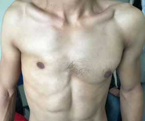
Absence of the chest muscle in one of our patients with Poland Syndrome
For the treatment of Poland Syndrome, see here for details.
Sternal Cleft
The sternum (breastbone) is formed by the midline fusion of two halves. When this fusion fails to occur because of a developmental disorder in the embryonic period, a sternal cleft is seen. The sternum may be completely cleft, or partially cleft depending on the point where fusion ends.
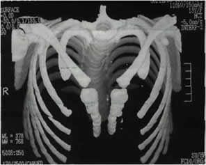
3D computed tomography image of a sternal cleft
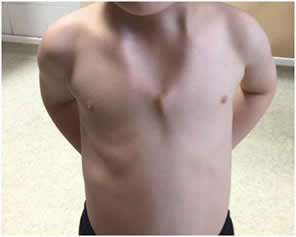
Image of a patient with a sternal cleft
Rib Flare
This is outward protrusion of the ribs. In this type of deformity, the patient is quite unhappy with the appearance. In the past, rib resection surgery was performed in all cases. Today, in addition to surgery, bandage (brace) therapy is also used to treat these deformities. In addition, with the Ribella Procedure, a painless, stitchless and scarless method developed and patented by Dr. Hasan Ersöz, patients can get rid of rib flare deformity with a very simple procedure in a short time. For details, you can visit the website of our Dr. Curver brand: DR CURVER website.